Multimedia Medical Conceptual Web for Intelligent Information Access |
| Scoliosis Scoliosis is a disease that causes deformations such as twisting and lateral bending of the spine. To correct scoliotic deformations by surgery and spinal fixation, the extents of 3D spinal deformations need to be measured. In principle, these measurements can be made on the 3D model reconstructed from the patient’s CT volume image of the spine. However, the radiation dosage of the patient in such a CT scan is too high. Therefore, x-ray imaging is currently the imaging technique of choice for the diagnosis and treatment of scoliosis. Multiple views of the patient’s spine can be taken at the same time using biplanar radiography or at different time using conventional radiography, with the patient turning to the side. Biplanar radiographic machines are bulky and inflexible. As they have limited use in clinical practice, they have been replaced by CT scanners. Therefore, conventional radiography is more commonly used for capturing x-ray images of scoliotic spines. Our method fits a model of the spine to that in the x-ray image to recover the 3D structure of the spine. Our first model is based on the Cosserat rod theory, which is able to model the bending and twisting of the spine. Sample Results 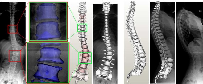 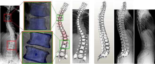 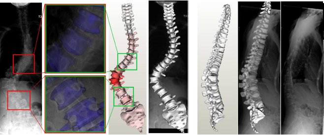 |
| Publications | |
| >>> | H. Li, W. K. Leow, C.-H. Huang, and T. S. Howe. Modeling and Measurement of 3D Deformations of Scoliotic Spine Using 2D X-ray Images. In Proc. Computer Analysis of Images and Patterns, pages 647-654, 2-4 Sep 2009. |
Last update: 30 Sep 2009