Automatic spinal stenosis detection
The Development of Magnetic Resonance Imaging (MRI) techniques has revolutionized our ability to see normal and abnormal spinal structures and help diagnose spine related diseases.
MRI spine is most commonly performed for a slipped disc. Patients are in a lot of pain and require rapid diagnosis. At NUH, from Jan to June in 2017, 3014 MRI spines were performed, while from Jan to June in 2019, 3360 MRI spines were performed, leading to an 11% increase. We need to report MRI spines timely and accurately.
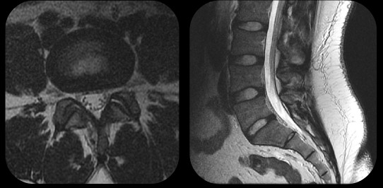
Figure 1: MRI spine studies performed on lumbar levels
In this project, we develop and apply AI techniques to automatically detect spine related diseases based on MRI images (three types of spinal stenosis): lateral stenosis, central stenosis, and foramina stenosis.
We propose a model pipeline for the spineAI project, which is shown in Figure 1 and Figure 2. Given an input spine MRI image, we first detect relevant spinal regions, and then label stenosis level with respective disease grades.
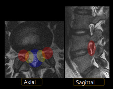
Figure 2: Automatic detection and grading of spinal stenosis at a lumbar level
This project is an on-going collaboration with NUHS Diagnostic imaging. 5 radiologists have been on board wih labeling and providing accurate visual data. Now our spine AI pipeline is detecting and grading narrowing of MRI spines at human level and comparable to junior radiologists.